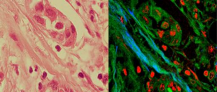
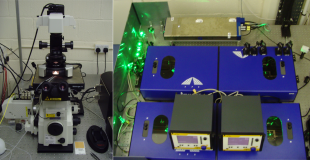
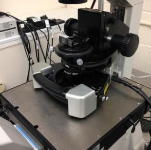
Biophotonics is the interface between Biology and Photonics that deals with the interaction between light and biological systems. The aim of the BioImaging Facility at Edinburgh University is to both investigate and exploit new imaging technologies and to provide access to state-of-the-art imaging instrumentation to users from biology and material sciences. Please review the listed equipment and services below for more information on how they can be of use in your research.
Confocal Raman Spectroscopy
What is Raman Spectroscopy?
Raman Spectroscopy provides a complete chemical fingerprint of biomolecules, cells and tissue and measures the quantity of each molecular bond present in the sample without the need for labels. Raman spectroscopy is a form of vibrational spectroscopy using visible or near-infrared light to characterise molecular compositions. This technique has three main advantages:
- The shorter wavelengths than IR spectroscopy enable measurement to a sub-cellular resolution with an improved water transmission. In addition, this allows the use of higher efficiency detectors.
- In addition, you can see some vibrations in the Raman spectrum that aren't visible in the infrared spectrum. (or example, Raman spectroscopy is superb for studying the carbon atoms)
- The laser power can be maintained at a level where the measurement can be considered non-invasive.
- In Raman spectroscopy, a laser is focussed onto a biomolecule. Some of this light loses energy in the interaction with the biomolecule and from this interaction has its wavelength-shifted. The distribution of the wavelength-shifted photons can then be measured on a spectrometer and used to identify the molecular bond responsible for shifting the wavelength based on the size of the shift.
What can Raman Spectroscopy show you?
The range of frequencies, or spectra, within molecules are well characterised. The Raman spectrum gives a fingerprint of the biochemical composition of the sample being measured. This spectra changes for the location in the cell marking the identifying the constituents under investigation.
What type of samples can be measured?
Generally, all materials in solid or liquid form produce Raman spectra, with the exception of pure metals. The signal strength is highest for pure crystalline materials; however, diluted samples can be easily measured by increasing the imaging duration. The confocal nature of the microscopes allows both surface and subsurface measurement, although only a few 10s of microns remain visible below the surface as the reduction in signal from attenuation limits the signal.
Label-Free Optical Microscopy
Label-free optical microscopy relies on scattering and absorption events to generate light from biological molecules with different wavelength without the need to stain or attach fluorescent proteins.
CARS (Coherent Anti-Stokes Raman Scattering)
CARS is a non-invasive microscopy that is able to image the chemical contrast of living biomolecules, cells and tissue within seconds.
- Uses the coherent anti-stoke transition to amplify the Raman signal to ~5 orders of magnitude higher
- Capable of providing fast imaging of living cells
- Easy implement other detection methods for colour images
SRS (Stimulated Raman Scattering)
Similar to CARS microscopy, SRS has grown in popularity because it has a reduced non-resonant background signal.
- Reduced non-resonant background signal
- SRS signal is proportional to the sample allowing quantitative measurement of contrast
- Identical spectrum to that of the Raman Spectroscopy for easier tuning
- Ability to measure lower concentrations than CARS
(TPEF) Two-Photon Excitation Fluorescence
TPEF is a multi-photon microscopy provided alongside CARS microscopy and is ideal for deep penetration within tissue and 3d sectioning without the risk of photo-toxicity or photo-bleaching. TPEF arises from the simultaneous absorption of two photons in a single quantised event.
- Uses both soft and hard aperture confocality to produce very thin sliced images
- Excitation in the near-IR provides greater penetration and imaging with white light
- Minimises the risk of photo-bleaching or photo-damage
SHG (Second Harmonic Generation)
SHG (also called "frequency doubling") is a nonlinear optical process, in which photons interacting with a nonlinear material are effectively "combined" to form new photons with twice the energy, and therefore twice the frequency and half the wavelength of the initial photons.
- Ideal for imaging nonlinear materials such as special chromophores or collagen fibres
- Retains optical properties of the input signal (coherent process)
- Measuring the intrinsic local second-order nonlinearity of a specimen
Atomic Force Microscopy
What is Atomic Force Microscopy
Atomic Force Microscopy is a method of quantitative imaging with a nanometre resolution comparable to that of electron microscopy. This microscopy provides images of both the topography and the biomechanical structure of biomolecules, cells and tissue.
What sort of information does AFM show?
- Resolution : Height < 0.1 nm Lateral < 20 nm (depends on tip radius)
- Can measure topography of diverse samples from:
- Live Cells, tissue and biomolecules
- Nanotubes or DNA strands
- Semiconductor Nano-structures
- Provides phase imaging for to allow quantitative imaging of sample mechanical/chemical properties
- No sample irradiation
Services Offered
- Access to both the multi-modal CARS/AFM microscope and Raman microscope
- Optimisation and calibration of the microscopes before use
- Training and supervision if needed using the device by experienced users
- Consultation as to ideal sample preparation
- Support with analysis and processing of images and measurements
- Support in Grant Application
Contact Name:
Contact Email:
Address:
Bioimaging Facility
Alrick Building
Kings Buildings
Max Born Crescent
EH9 3BF
United Kingdom


